A bioengineer, Louise Thines completed her dissertation in molecular and cellular biology at UCLouvain before continuing her research for three and a half years in Washington, D.C., in the United States. She then joined the University of Namur for a new postdoctoral fellowship in Professor Henri-François Renard’s laboratory, with support from MSCA funding.

"Velcro" on the surface of the cells?

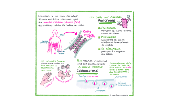
In tissues, cells are constantly interacting with their neighbors and their environment. Certain proteins present in the cell membrane play a direct role in these interactions. This is the case with cell adhesion molecules, often referred to by the acronym CAMs.

These CAMs can be compared to tiny molecular Velcro fasteners that help keep cells in contact. They also play a role in transmitting signals into the cell and can influence the cell’s organization, movement, or interactions with neighboring cells. To control these interactions, the cell can add certain proteins to its membrane, move them, recycle them, or remove them. One of the mechanisms used to bring them into the cell is endocytosis.

CAMs play an important role in the ability of tissues to form, renew themselves, and repair themselves. During embryonic development or wound healing, for example, certain cells must modify their contacts with neighboring cells in order to change position and participate in tissue remodeling. By adjusting the adhesion molecules on their surfaces, they can strengthen certain contacts, loosen others, and become more mobile. This ability to modify intercellular adhesions is essential for the body’s normal functioning.

Facilitation visuelle - CAMS

From Phagocytosis to Endocytosis

The process by which a cell takes in material (phagocytosis) was first described in the late 19th century by Élie Metchnikoff, a Russian zoologist working at the Pasteur Institute. Throughout the 20th century, extensive research led to the definition of the concept of endocytosis, which refers to the set of mechanisms involved in the internalization of particles. Today, research focuses on the molecular mechanisms underlying this phenomenon. A better understanding of a mechanism opens the door to developing tools or methods to control it for medical and pharmaceutical purposes.

Several entry points

During the process of endocytosis, the cell membrane bulges around a molecule on its surface. It gradually forms a pit that eventually closes off inside the cell, taking the form of a small vesicle. 

Endocytosis can occur through several “pathways.” It’s a bit like a building with several types of doors operated by mechanisms that use different technologies. 

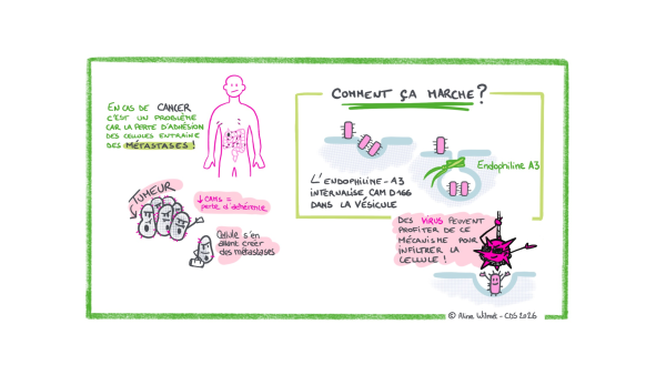
The best-characterized pathway today involves a protein called clathrin, which, thanks to its structure, causes the cell membrane to curve inward until a vesicle is formed. But not all molecules follow this route.In 2020, research by Henri-François Renard revealed another endocytosis pathway. This pathway operates independently of clathrin and involves, in particular, endophilin-A3 to facilitate the entry of adhesion molecules into the cell.

It is this mechanism—which is still not fully understood—that lies at the heart of Louise’s project.

Why is this so important? Because in certain cancers, tumor cells can lose their ability to adhere, gain greater migratory capacity, and invade neighboring tissues. This process contributes to the various stages that can lead to the formation of metastases. Understanding how CAMs are removed or maintained on the cell surface therefore also provides insight into certain mechanisms associated with tumor progression. Furthermore, certain viruses can use CAMs as “Velcro” by attaching to them to gain entry into our cells. Targeting the CAM entry point could therefore be a promising strategy for combating certain viral diseases.

Facilitation visuelle - Cancer

The ModulEndoA3 Project: Understanding How Cells Control Their Adhesion Sites

Louise is trying to determine how this endocytosis pathway works at the molecular level. Which molecules are involved? How do they interact? What triggers the internalization of an adhesion molecule into the cell? What happens when one of the components involved in this mechanism is disrupted?

In particular, Louise is studying the “Velcro” protein CD166, a cell adhesion molecule (CAM) found on the surface of various cells.

Observe the cells to take the machinery apart

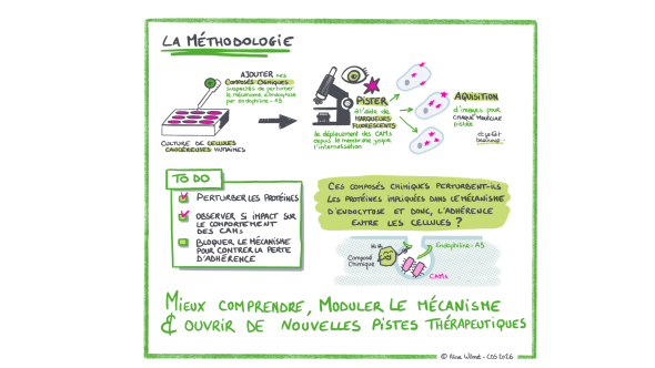
To understand what is happening, Louise is working with human cancer cells grown in the laboratory.

She can modify the activity or abundance of proteins suspected of being involved in the mechanism, using genetic tools or chemical compounds. She then observes the consequences of these disruptions.

Microscopy plays a central role here. Using fluorescent markers, it becomes possible to visualize different proteins within the cells and track their location. Louise can thus simultaneously observe endophilin-A3 and the adhesion molecules that have been internalized into the cell’s cytoplasm.

The images serve not only to illustrate the experiment. They become data that the researcher can quantify and compare to determine whether an experimental modification accelerates, slows down, or disrupts endocytosis.

Hundreds of molecules were screened

To better understand the mechanisms of this endocytosis pathway, Louise is also seeking to identify other proteins that may be involved in its function.

In collaboration with the University of Geneva, she is testing hundreds of chemical compounds capable of affecting various proteins within the cell. After each treatment, images are automatically captured under a microscope to observe whether the endocytosis process is altered. Most of the compounds tested will likely have no particular effect. But if one of them significantly slows down or disrupts the mechanism, it is the protein it targets that becomes particularly interesting: it could be involved, directly or indirectly, in this endocytosis pathway. Further analyses are then needed to verify its role. These compounds are thus valuable tools for research. By blocking a protein and observing the consequences on the cell, researchers can gradually understand its function.

Certain molecules capable of modulating this pathway could also, in the longer term, open up avenues for new therapeutic strategies. At this stage, however, these are not yet drugs: between the identification of an active compound in cell culture and the development of a treatment, many research steps remain necessary.

Broadening Our Understanding of Endocytosis

Beyond its potential applications, Louise Thines’s project touches on a fundamental question in cell biology.

Traditional models have long been used to describe a few major pathways that cells use to internalize molecules on their surface. The discovery and study of clathrin-independent mechanisms now reveal a more diverse landscape. Understanding this diversity provides deeper insight into how a cell continuously regulates its interface with its environment and adapts its behavior.

The MSCA funding enables Louise to continue this exploration, develop new experimental approaches, and strengthen international collaborations. The results are expected, in particular, to help clarify the functioning of this endocytosis pathway and to identify new molecules capable of modulating it.

The Marie Skłodowska-Curie Fellowship (MSCA-PF)

The Marie Skłodowska-Curie Fellowship (MSCA-PF) is a selective European grant that enables a postdoctoral researcher to carry out an ambitious project at a new institution, often abroad. It supports the excellence of the project, the researcher’s mobility, and the development of their scientific career.

Logo "Financé par l'Union européenne"

European Researchers' Night

To mark European Researchers’ Night, UNamur is highlighting several research projects conducted as part of the Marie Skłodowska-Curie Actions (MSCA). To make this research and its implications accessible to as many people as possible, Confluent des Savoirs—UNamur’s research outreach and dissemination service—has created a visual summary of each project. These visualizations aim to organize scientific information into illustrated, accessible summaries that combine keywords, diagrams, and graphics to help readers quickly grasp the mechanisms being studied and the key research questions.

These projects are part of the European Researchers’ Night initiative, which each year invites the public to discover research, the people behind it, and its role in our understanding of the world.