The Biology Department strives to understand the living world in all its diversity, in the course of its evolution, from molecules to ecosystems, and to respond to complex societal problems such as biodiversity loss, infectious diseases, antibiotic resistance, etc. Biologists use an approach that integrates the laboratory, the field and computer tools.

Find out more about the department

SVG

Studies

See content
SVG

Research

See content
SVG

Organization chart

See content
SVG

Alumni

See content
SVG

Contact

See content

Biology is a modern science on the move. It responds to complex societal problems such as biodiversity loss, antibiotic resistance, adaptation to climate change, the fight against chronic or infectious diseases, food safety... In short, biology is present everywhere in society.

Biology uses a wide range of scientific and technological knowledge and approaches that enable it to address the living world in all its diversity (viruses, bacteria, fungi, plants, parasites, animals, humans), throughout its evolution and at all levels of organization (molecules, cells, tissues, individuals, super-organisms, populations, ecosystems).

From knowledge of fundamental processes to multiple applications, the 21st century biologist is a complete scientist, wielding the scientific approach, as much in the laboratory as when out in the field (in natural areas) or when using computer analysis methods (and processing high-throughput data).
Spotlight

News

Cellular "Velcro" Under Control

Biology
Life and health sciences
Outreach

How does the study of a cellular entry point shed light on certain mechanisms of cancer and viral infections?

Our skin, the lining of our intestines, and even our blood vessels are made up of cells organized into tissues. To stay in contact, communicate, and adapt their behavior, these cells rely in particular on proteins present on their surface. Among these, cell adhesion molecules play an essential role. Louise Thines, a researcher at the University of Namur and recipient of a Marie Skłodowska-Curie Actions (MSCA) grant, is studying how some of these proteins are internalized by cells, thereby reducing adhesion between them. This approach to understanding the mechanism offers avenues of research that are also relevant to the fight against cancer and viral infections.
Facilitation visuelle - Méthodologie

A bioengineer, Louise Thines completed her dissertation in molecular and cellular biology at UCLouvain before continuing her research for three and a half years in Washington, D.C., in the United States. She then joined the University of Namur for a new postdoctoral fellowship in Professor Henri-François Renard’s laboratory, with support from MSCA funding.

"Velcro" on the surface of the cells?

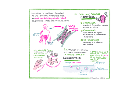
In tissues, cells are constantly interacting with their neighbors and their environment. Certain proteins present in the cell membrane play a direct role in these interactions. This is the case with cell adhesion molecules, often referred to by the acronym CAMs.

These CAMs can be compared to tiny molecular Velcro fasteners that help keep cells in contact. They also play a role in transmitting signals into the cell and can influence the cell’s organization, movement, or interactions with neighboring cells. To control these interactions, the cell can add certain proteins to its membrane, move them, recycle them, or remove them. One of the mechanisms used to bring them into the cell is endocytosis.

CAMs play an important role in the ability of tissues to form, renew themselves, and repair themselves. During embryonic development or wound healing, for example, certain cells must modify their contacts with neighboring cells in order to change position and participate in tissue remodeling. By adjusting the adhesion molecules on their surfaces, they can strengthen certain contacts, loosen others, and become more mobile. This ability to modify intercellular adhesions is essential for the body’s normal functioning.
Facilitation visuelle - CAMS

From Phagocytosis to Endocytosis

The process by which a cell takes in material (phagocytosis) was first described in the late 19th century by Élie Metchnikoff, a Russian zoologist working at the Pasteur Institute. Throughout the 20th century, extensive research led to the definition of the concept of endocytosis, which refers to the set of mechanisms involved in the internalization of particles. Today, research focuses on the molecular mechanisms underlying this phenomenon. A better understanding of a mechanism opens the door to developing tools or methods to control it for medical and pharmaceutical purposes.

Several entry points

During the process of endocytosis, the cell membrane bulges around a molecule on its surface. It gradually forms a pit that eventually closes off inside the cell, taking the form of a small vesicle. 

Endocytosis can occur through several “pathways.” It’s a bit like a building with several types of doors operated by mechanisms that use different technologies. 

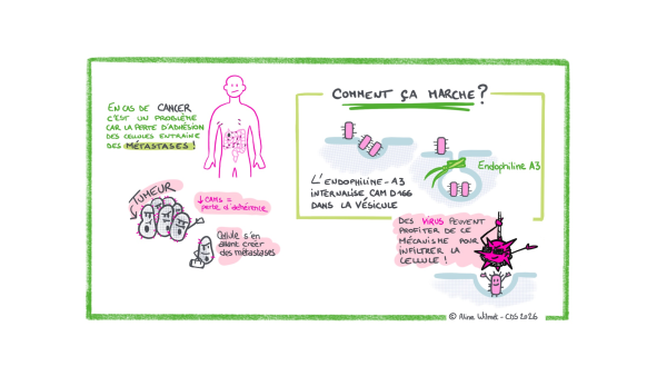
The best-characterized pathway today involves a protein called clathrin, which, thanks to its structure, causes the cell membrane to curve inward until a vesicle is formed. But not all molecules follow this route.In 2020, research by Henri-François Renard revealed another endocytosis pathway. This pathway operates independently of clathrin and involves, in particular, endophilin-A3 to facilitate the entry of adhesion molecules into the cell.

It is this mechanism—which is still not fully understood—that lies at the heart of Louise’s project.

Why is this so important? Because in certain cancers, tumor cells can lose their ability to adhere, gain greater migratory capacity, and invade neighboring tissues. This process contributes to the various stages that can lead to the formation of metastases. Understanding how CAMs are removed or maintained on the cell surface therefore also provides insight into certain mechanisms associated with tumor progression. Furthermore, certain viruses can use CAMs as “Velcro” by attaching to them to gain entry into our cells. Targeting the CAM entry point could therefore be a promising strategy for combating certain viral diseases.
Facilitation visuelle - Cancer

The ModulEndoA3 Project: Understanding How Cells Control Their Adhesion Sites

Louise is trying to determine how this endocytosis pathway works at the molecular level. Which molecules are involved? How do they interact? What triggers the internalization of an adhesion molecule into the cell? What happens when one of the components involved in this mechanism is disrupted?

In particular, Louise is studying the “Velcro” protein CD166, a cell adhesion molecule (CAM) found on the surface of various cells.

Observe the cells to take the machinery apart

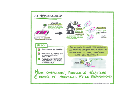
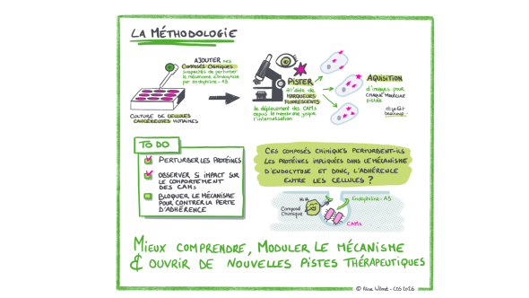
To understand what is happening, Louise is working with human cancer cells grown in the laboratory.

She can modify the activity or abundance of proteins suspected of being involved in the mechanism, using genetic tools or chemical compounds. She then observes the consequences of these disruptions.

Microscopy plays a central role here. Using fluorescent markers, it becomes possible to visualize different proteins within the cells and track their location. Louise can thus simultaneously observe endophilin-A3 and the adhesion molecules that have been internalized into the cell’s cytoplasm.

The images serve not only to illustrate the experiment. They become data that the researcher can quantify and compare to determine whether an experimental modification accelerates, slows down, or disrupts endocytosis.

Hundreds of molecules were screened

To better understand the mechanisms of this endocytosis pathway, Louise is also seeking to identify other proteins that may be involved in its function.

In collaboration with the University of Geneva, she is testing hundreds of chemical compounds capable of affecting various proteins within the cell. After each treatment, images are automatically captured under a microscope to observe whether the endocytosis process is altered. Most of the compounds tested will likely have no particular effect. But if one of them significantly slows down or disrupts the mechanism, it is the protein it targets that becomes particularly interesting: it could be involved, directly or indirectly, in this endocytosis pathway. Further analyses are then needed to verify its role. These compounds are thus valuable tools for research. By blocking a protein and observing the consequences on the cell, researchers can gradually understand its function.

Certain molecules capable of modulating this pathway could also, in the longer term, open up avenues for new therapeutic strategies. At this stage, however, these are not yet drugs: between the identification of an active compound in cell culture and the development of a treatment, many research steps remain necessary.

Broadening Our Understanding of Endocytosis

Beyond its potential applications, Louise Thines’s project touches on a fundamental question in cell biology.

Traditional models have long been used to describe a few major pathways that cells use to internalize molecules on their surface. The discovery and study of clathrin-independent mechanisms now reveal a more diverse landscape. Understanding this diversity provides deeper insight into how a cell continuously regulates its interface with its environment and adapts its behavior.

The MSCA funding enables Louise to continue this exploration, develop new experimental approaches, and strengthen international collaborations. The results are expected, in particular, to help clarify the functioning of this endocytosis pathway and to identify new molecules capable of modulating it.

The Marie Skłodowska-Curie Fellowship (MSCA-PF)

The Marie Skłodowska-Curie Fellowship (MSCA-PF) is a selective European grant that enables a postdoctoral researcher to carry out an ambitious project at a new institution, often abroad. It supports the excellence of the project, the researcher’s mobility, and the development of their scientific career.

Logo "Financé par l'Union européenne"

European Researchers' Night

To mark European Researchers’ Night, UNamur is highlighting several research projects conducted as part of the Marie Skłodowska-Curie Actions (MSCA). To make this research and its implications accessible to as many people as possible, Confluent des Savoirs—UNamur’s research outreach and dissemination service—has created a visual summary of each project. These visualizations aim to organize scientific information into illustrated, accessible summaries that combine keywords, diagrams, and graphics to help readers quickly grasp the mechanisms being studied and the key research questions.

These projects are part of the European Researchers’ Night initiative, which each year invites the public to discover research, the people behind it, and its role in our understanding of the world.
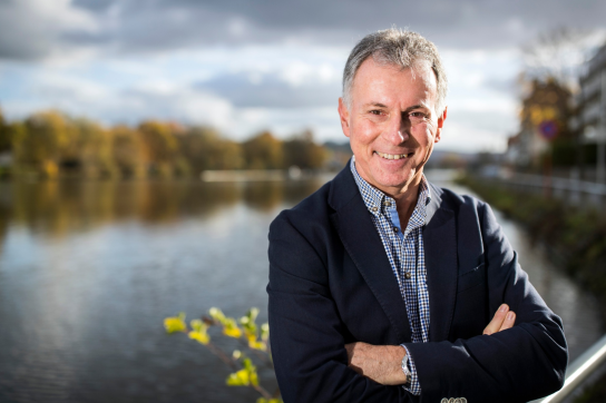
Through the work of Patrick Kestemont, UNamur is contributing to the captive breeding of a catfish adapted to the saltwater of the Mekong River

Biodiversity

After more than two years of research, a team comprising the University of Namur, the University of Liège, and Can Tho University in Vietnam has succeeded in breeding Pangasius krempfi in captivity—a catfish capable of living in both freshwater and brackish water. This is a significant step forward for the DIVERSAQUA project, which aims to develop aquaculture better suited to the increasing salinization of the Mekong Delta. 
Patrick Kestemont
Félicitations à l’ensemble des chercheurs impliqués dans le projet DIVERSAQUA, et tout particulièrement au professeur Patrick Kestemont, qui coordonne le projet pour l’UNamur !

In the Mekong Delta, aquaculture is increasingly facing the encroachment of saltwater into areas that are typically supplied with freshwater. This phenomenon, known as salinity intrusion, can make it more difficult to farm certain locally raised species. 

It is against this backdrop that the DIVERSAQUA project is being carried out, funded by ARES-CCI and coordinated by Professor Patrick Kestemont of UNamur and Professor Nguyen Thanh Phuong of Can Tho University. The project also involves the University of Liège. Its goal is to identify and better manage the farming of species capable of withstanding these changes in salinity. 

A fish capable of adapting to various levels of salinity

The researchers focused on Pangasius krempfi, known locally as ca bong lau or ca dua. This catfish is considered euryhaline, meaning it can live in waters with varying salinity levels. This characteristic makes it a particularly promising candidate for areas of the Mekong Delta that are increasingly affected by saltwater intrusion. 

The species also has other advantages: it can grow to a large size, its flesh is highly prized, and it has significant economic value. In the delta, it is found primarily in estuaries and coastal areas. Until now, however, its aquaculture has remained limited.  

The first successful breeding in captivity

After more than two years of work, the project teams succeeded in raising wild juvenile fish to adulthood in a closed-loop aquaculture system, where the water is filtered and reused. The mature fish were then induced to reproduce using hormones commonly used in aquaculture. The resulting larvae were recently reared in tanks until they reached the juvenile stage. Eventually, these animals could become the first fully domesticated broodstock of the species.

The goal is to gradually produce the juvenile fish needed for aquaculture without relying solely on wild populations. This would ensure a more consistent supply and allow for continued research into the commercial farming of the species. 

Gaining a Better Understanding of Salt Tolerance

At the same time, researchers are also studying Pangasius krempfi’s ability to tolerate different salt concentrations. Several experiments are being conducted at various stages of its development—from the larval stage to the juvenile stage—to better understand its physiological responses and immune system. 

This research should help determine the conditions under which the species can be farmed and more accurately assess its potential in areas subject to saltwater intrusion. In the longer term, the DIVERSAQUA project aims to diversify the species farmed in the Mekong Delta and offer new solutions to aquaculturists facing the effects of climate change. The successful reproduction of Pangasius krempfi in captivity is therefore an important first step in this direction. The teams are now continuing their research with the goal of gaining a better understanding of the species and assessing the conditions necessary for its development on a larger scale. Congratulations to all the researchers involved in the DIVERSAQUA project, and especially to Professor Patrick Kestemont, who coordinates the project for UNamur!  
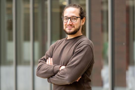
A paper published in *Nature Human Behavior* highlights the barriers to creativity among young researchers

Publication

Published in *Nature Human Behaviour*, an international study to which Eli Thoré—a professor at UNamur and a member of the Institute of Life-Earth-Environment—contributed examines the conditions necessary to foster greater scientific creativity. He discusses this project and what he has learned from it.
Eli Thoré
Eli Thoré, professeur assistant à l’UNamur et coauteur de l’étude publiée dans Nature Human Behaviour.

Pressure to publish, short-term funding, competition, and the precarious nature of early-career stages: Can these constraints push young researchers to prioritize “safer” projects at the expense of more original ideas? This is the central question of an international publication to which Eli Thoré, an assistant professor at the University of Namur, contributed.

Having been interested for several years in “metascience”—that is, the study of how science itself works—he participated in the discussions, the development of ideas, and the drafting and revision of the manuscript. The project, launched at the initiative of Phillip Haubrock in late 2025, led to numerous exchanges among the authors before it was finally published in *Nature Human Behaviour*. “The exchanges were very fruitful and, at first, quite chaotic!” says Eli Thoré.

Constraints that stifle creativity

To conduct their analysis, the authors compiled numerous studies on the workings of the academic world. Their conclusion is clear: scientific creativity is not merely an individual trait; it also depends on the conditions under which researchers work. 

Short-term funding, pressure to publish, emphasis on the number of articles or the prestige of journals, fierce competition for positions or funding… All these constraints can push researchers to prioritize projects whose results are easier to predict, at the expense of more original or risky ideas. 

This situation particularly affects early-career researchers. They often have less autonomy, and their future depends more heavily on upcoming publications, grant applications, or job opportunities. Taking a risk in one’s research can thus also become a risk to one’s career. 

Image
Eli Thoré

“The problem isn’t that young researchers lack creativity, but that the system doesn’t always provide them with the conditions they need to express it.”

Eli Thoré Teaching Assistant at UNamur

The consequences can be significant. For example, a study cited in the publication shows that narrowly failing to secure key funding early in one’s career increases the risk of leaving academia by more than 10 percent. 

Eli Thoré himself acknowledges having felt this tension between creativity and the need to demonstrate in advance that a project will succeed. During his Ph.D., he became interested in the daily rhythms of animal behavior—similar to our sleep-wake cycle—and how they might modify the effects of environmental pollution. At the time, this line of inquiry was quite removed from his main research project.  Yet he decided to pursue it. Over time, this idea became a central focus of his research. “If I had limited myself to questions I already knew were safe and easy to justify, I might never have gotten here,” he explains.

More time, stability, and autonomy

The authors propose several approaches to create an environment more conducive to this kind of scientific risk-taking. These include: providing funding over longer periods, offering greater stability to early-career researchers, reducing certain administrative burdens, and revising the criteria used to evaluate a scientific career. 

The idea, in particular, is to no longer measure success solely through easily quantifiable indicators, such as the number of publications or the prestige of journals, but to give greater weight to originality, collaboration, perseverance, and scientific independence. The publication also emphasizes three essential conditions for creativity: time, autonomy, and the freedom to fail.

Image
Eli Thoré

“Some of the most interesting discoveries probably begin with an idea that at first seems strange, uncertain, or even a little crazy.”

Eli Thoré Teaching Assistant at UNamur

The paper therefore puts forward an idea that seems fairly simple on the surface: if we want researchers to be creative, we must give them the freedom to be so. This is a particularly important issue for young scientists, who will shape the future directions of research. Congratulations to Eli Thoré and all the researchers involved in this work on their publication in *Nature Human Behaviour*! 

Researchers from Namur Achieve Great Success in the F.R.S.-FNRS’s 2026 “Grants and Research Awards” and “Télévie” Calls

Institution

On June 23, 2026, the F.R.S.-FNRS published the list of recipients of various doctoral and postdoctoral fellowships and Télévie projects (cancer-focused research). Among them, numerous researchers from UNamur received funding.
Logo FNRS

Six researchers have been awarded doctoral fellowships to begin their doctoral dissertations: Rachel LAURON from the Faculty of Sciences; Océane WATELET, Vera NOVAK, Camille LAMBIET, Alionka WÉRENNE, and Théodore HARDY (who received his grant from ULB under a joint supervision arrangement with UNamur) from the Faculty of Philosophy and Letters. 

Researchers from Namur also achieved great success in securing research fellow grants. Seven of them received this postdoctoral funding. They are Eleonor CELORA, Nataliya PUCHENKINA, Jérémy ARTRU and Bernardino PITOCCHELLI from the Faculty of Philosophy and Letters; Romain MERTENS from the Faculty of Law; and David TALUKDER and François WOITRIN from the Faculty of Economics, Management, and Communication at SciencesPo (EMCP). 

In addition, two permanent F.R.S.-FNRS researchers at UNamur have been promoted to Senior Researcher: Francesca CECCHET and Yves CAUDANO, both members of the Department of Physics and the NISM Institute. 

The Télévie call for proposals also enabled Marc HENNEQUART to secure funding to begin research aimed at identifying the metabolic determinants of the response to arginine deprivation in pancreatic and colorectal cancers. 

Congratulations to them!


Cellular "Velcro" Under Control

Biology
Life and health sciences
Outreach

How does the study of a cellular entry point shed light on certain mechanisms of cancer and viral infections?

Our skin, the lining of our intestines, and even our blood vessels are made up of cells organized into tissues. To stay in contact, communicate, and adapt their behavior, these cells rely in particular on proteins present on their surface. Among these, cell adhesion molecules play an essential role. Louise Thines, a researcher at the University of Namur and recipient of a Marie Skłodowska-Curie Actions (MSCA) grant, is studying how some of these proteins are internalized by cells, thereby reducing adhesion between them. This approach to understanding the mechanism offers avenues of research that are also relevant to the fight against cancer and viral infections.

Facilitation visuelle - Méthodologie

A bioengineer, Louise Thines completed her dissertation in molecular and cellular biology at UCLouvain before continuing her research for three and a half years in Washington, D.C., in the United States. She then joined the University of Namur for a new postdoctoral fellowship in Professor Henri-François Renard’s laboratory, with support from MSCA funding.

"Velcro" on the surface of the cells?

In tissues, cells are constantly interacting with their neighbors and their environment. Certain proteins present in the cell membrane play a direct role in these interactions. This is the case with cell adhesion molecules, often referred to by the acronym CAMs.

These CAMs can be compared to tiny molecular Velcro fasteners that help keep cells in contact. They also play a role in transmitting signals into the cell and can influence the cell’s organization, movement, or interactions with neighboring cells. To control these interactions, the cell can add certain proteins to its membrane, move them, recycle them, or remove them. One of the mechanisms used to bring them into the cell is endocytosis.

CAMs play an important role in the ability of tissues to form, renew themselves, and repair themselves. During embryonic development or wound healing, for example, certain cells must modify their contacts with neighboring cells in order to change position and participate in tissue remodeling. By adjusting the adhesion molecules on their surfaces, they can strengthen certain contacts, loosen others, and become more mobile. This ability to modify intercellular adhesions is essential for the body’s normal functioning.

Facilitation visuelle - CAMS

From Phagocytosis to Endocytosis

The process by which a cell takes in material (phagocytosis) was first described in the late 19th century by Élie Metchnikoff, a Russian zoologist working at the Pasteur Institute. Throughout the 20th century, extensive research led to the definition of the concept of endocytosis, which refers to the set of mechanisms involved in the internalization of particles. Today, research focuses on the molecular mechanisms underlying this phenomenon. A better understanding of a mechanism opens the door to developing tools or methods to control it for medical and pharmaceutical purposes.

Several entry points

During the process of endocytosis, the cell membrane bulges around a molecule on its surface. It gradually forms a pit that eventually closes off inside the cell, taking the form of a small vesicle. 

Endocytosis can occur through several “pathways.” It’s a bit like a building with several types of doors operated by mechanisms that use different technologies. 

The best-characterized pathway today involves a protein called clathrin, which, thanks to its structure, causes the cell membrane to curve inward until a vesicle is formed. But not all molecules follow this route.In 2020, research by Henri-François Renard revealed another endocytosis pathway. This pathway operates independently of clathrin and involves, in particular, endophilin-A3 to facilitate the entry of adhesion molecules into the cell.

It is this mechanism—which is still not fully understood—that lies at the heart of Louise’s project.

Why is this so important? Because in certain cancers, tumor cells can lose their ability to adhere, gain greater migratory capacity, and invade neighboring tissues. This process contributes to the various stages that can lead to the formation of metastases. Understanding how CAMs are removed or maintained on the cell surface therefore also provides insight into certain mechanisms associated with tumor progression. Furthermore, certain viruses can use CAMs as “Velcro” by attaching to them to gain entry into our cells. Targeting the CAM entry point could therefore be a promising strategy for combating certain viral diseases.

Facilitation visuelle - Cancer

The ModulEndoA3 Project: Understanding How Cells Control Their Adhesion Sites

Louise is trying to determine how this endocytosis pathway works at the molecular level. Which molecules are involved? How do they interact? What triggers the internalization of an adhesion molecule into the cell? What happens when one of the components involved in this mechanism is disrupted?

In particular, Louise is studying the “Velcro” protein CD166, a cell adhesion molecule (CAM) found on the surface of various cells.

Observe the cells to take the machinery apart

To understand what is happening, Louise is working with human cancer cells grown in the laboratory.

She can modify the activity or abundance of proteins suspected of being involved in the mechanism, using genetic tools or chemical compounds. She then observes the consequences of these disruptions.

Microscopy plays a central role here. Using fluorescent markers, it becomes possible to visualize different proteins within the cells and track their location. Louise can thus simultaneously observe endophilin-A3 and the adhesion molecules that have been internalized into the cell’s cytoplasm.

The images serve not only to illustrate the experiment. They become data that the researcher can quantify and compare to determine whether an experimental modification accelerates, slows down, or disrupts endocytosis.

Hundreds of molecules were screened

To better understand the mechanisms of this endocytosis pathway, Louise is also seeking to identify other proteins that may be involved in its function.

In collaboration with the University of Geneva, she is testing hundreds of chemical compounds capable of affecting various proteins within the cell. After each treatment, images are automatically captured under a microscope to observe whether the endocytosis process is altered. Most of the compounds tested will likely have no particular effect. But if one of them significantly slows down or disrupts the mechanism, it is the protein it targets that becomes particularly interesting: it could be involved, directly or indirectly, in this endocytosis pathway. Further analyses are then needed to verify its role. These compounds are thus valuable tools for research. By blocking a protein and observing the consequences on the cell, researchers can gradually understand its function.

Certain molecules capable of modulating this pathway could also, in the longer term, open up avenues for new therapeutic strategies. At this stage, however, these are not yet drugs: between the identification of an active compound in cell culture and the development of a treatment, many research steps remain necessary.

Broadening Our Understanding of Endocytosis

Beyond its potential applications, Louise Thines’s project touches on a fundamental question in cell biology.

Traditional models have long been used to describe a few major pathways that cells use to internalize molecules on their surface. The discovery and study of clathrin-independent mechanisms now reveal a more diverse landscape. Understanding this diversity provides deeper insight into how a cell continuously regulates its interface with its environment and adapts its behavior.

The MSCA funding enables Louise to continue this exploration, develop new experimental approaches, and strengthen international collaborations. The results are expected, in particular, to help clarify the functioning of this endocytosis pathway and to identify new molecules capable of modulating it.

The Marie Skłodowska-Curie Fellowship (MSCA-PF)

The Marie Skłodowska-Curie Fellowship (MSCA-PF) is a selective European grant that enables a postdoctoral researcher to carry out an ambitious project at a new institution, often abroad. It supports the excellence of the project, the researcher’s mobility, and the development of their scientific career.

Logo "Financé par l'Union européenne"

European Researchers' Night

To mark European Researchers’ Night, UNamur is highlighting several research projects conducted as part of the Marie Skłodowska-Curie Actions (MSCA). To make this research and its implications accessible to as many people as possible, Confluent des Savoirs—UNamur’s research outreach and dissemination service—has created a visual summary of each project. These visualizations aim to organize scientific information into illustrated, accessible summaries that combine keywords, diagrams, and graphics to help readers quickly grasp the mechanisms being studied and the key research questions.

These projects are part of the European Researchers’ Night initiative, which each year invites the public to discover research, the people behind it, and its role in our understanding of the world.

Through the work of Patrick Kestemont, UNamur is contributing to the captive breeding of a catfish adapted to the saltwater of the Mekong River

Biodiversity

After more than two years of research, a team comprising the University of Namur, the University of Liège, and Can Tho University in Vietnam has succeeded in breeding Pangasius krempfi in captivity—a catfish capable of living in both freshwater and brackish water. This is a significant step forward for the DIVERSAQUA project, which aims to develop aquaculture better suited to the increasing salinization of the Mekong Delta. 

Patrick Kestemont
Félicitations à l’ensemble des chercheurs impliqués dans le projet DIVERSAQUA, et tout particulièrement au professeur Patrick Kestemont, qui coordonne le projet pour l’UNamur !

In the Mekong Delta, aquaculture is increasingly facing the encroachment of saltwater into areas that are typically supplied with freshwater. This phenomenon, known as salinity intrusion, can make it more difficult to farm certain locally raised species. 

It is against this backdrop that the DIVERSAQUA project is being carried out, funded by ARES-CCI and coordinated by Professor Patrick Kestemont of UNamur and Professor Nguyen Thanh Phuong of Can Tho University. The project also involves the University of Liège. Its goal is to identify and better manage the farming of species capable of withstanding these changes in salinity. 

A fish capable of adapting to various levels of salinity

The researchers focused on Pangasius krempfi, known locally as ca bong lau or ca dua. This catfish is considered euryhaline, meaning it can live in waters with varying salinity levels. This characteristic makes it a particularly promising candidate for areas of the Mekong Delta that are increasingly affected by saltwater intrusion. 

The species also has other advantages: it can grow to a large size, its flesh is highly prized, and it has significant economic value. In the delta, it is found primarily in estuaries and coastal areas. Until now, however, its aquaculture has remained limited.  

The first successful breeding in captivity

After more than two years of work, the project teams succeeded in raising wild juvenile fish to adulthood in a closed-loop aquaculture system, where the water is filtered and reused. The mature fish were then induced to reproduce using hormones commonly used in aquaculture. The resulting larvae were recently reared in tanks until they reached the juvenile stage. Eventually, these animals could become the first fully domesticated broodstock of the species.

The goal is to gradually produce the juvenile fish needed for aquaculture without relying solely on wild populations. This would ensure a more consistent supply and allow for continued research into the commercial farming of the species. 

Gaining a Better Understanding of Salt Tolerance

At the same time, researchers are also studying Pangasius krempfi’s ability to tolerate different salt concentrations. Several experiments are being conducted at various stages of its development—from the larval stage to the juvenile stage—to better understand its physiological responses and immune system. 

This research should help determine the conditions under which the species can be farmed and more accurately assess its potential in areas subject to saltwater intrusion. In the longer term, the DIVERSAQUA project aims to diversify the species farmed in the Mekong Delta and offer new solutions to aquaculturists facing the effects of climate change. The successful reproduction of Pangasius krempfi in captivity is therefore an important first step in this direction. The teams are now continuing their research with the goal of gaining a better understanding of the species and assessing the conditions necessary for its development on a larger scale. Congratulations to all the researchers involved in the DIVERSAQUA project, and especially to Professor Patrick Kestemont, who coordinates the project for UNamur!  

A paper published in *Nature Human Behavior* highlights the barriers to creativity among young researchers

Publication

Published in *Nature Human Behaviour*, an international study to which Eli Thoré—a professor at UNamur and a member of the Institute of Life-Earth-Environment—contributed examines the conditions necessary to foster greater scientific creativity. He discusses this project and what he has learned from it.

Eli Thoré
Eli Thoré, professeur assistant à l’UNamur et coauteur de l’étude publiée dans Nature Human Behaviour.

Pressure to publish, short-term funding, competition, and the precarious nature of early-career stages: Can these constraints push young researchers to prioritize “safer” projects at the expense of more original ideas? This is the central question of an international publication to which Eli Thoré, an assistant professor at the University of Namur, contributed.

Having been interested for several years in “metascience”—that is, the study of how science itself works—he participated in the discussions, the development of ideas, and the drafting and revision of the manuscript. The project, launched at the initiative of Phillip Haubrock in late 2025, led to numerous exchanges among the authors before it was finally published in *Nature Human Behaviour*. “The exchanges were very fruitful and, at first, quite chaotic!” says Eli Thoré.

Constraints that stifle creativity

To conduct their analysis, the authors compiled numerous studies on the workings of the academic world. Their conclusion is clear: scientific creativity is not merely an individual trait; it also depends on the conditions under which researchers work. 

Short-term funding, pressure to publish, emphasis on the number of articles or the prestige of journals, fierce competition for positions or funding… All these constraints can push researchers to prioritize projects whose results are easier to predict, at the expense of more original or risky ideas. 

This situation particularly affects early-career researchers. They often have less autonomy, and their future depends more heavily on upcoming publications, grant applications, or job opportunities. Taking a risk in one’s research can thus also become a risk to one’s career. 

Image
Eli Thoré

“The problem isn’t that young researchers lack creativity, but that the system doesn’t always provide them with the conditions they need to express it.”

Eli Thoré Teaching Assistant at UNamur

The consequences can be significant. For example, a study cited in the publication shows that narrowly failing to secure key funding early in one’s career increases the risk of leaving academia by more than 10 percent. 

Eli Thoré himself acknowledges having felt this tension between creativity and the need to demonstrate in advance that a project will succeed. During his Ph.D., he became interested in the daily rhythms of animal behavior—similar to our sleep-wake cycle—and how they might modify the effects of environmental pollution. At the time, this line of inquiry was quite removed from his main research project.  Yet he decided to pursue it. Over time, this idea became a central focus of his research. “If I had limited myself to questions I already knew were safe and easy to justify, I might never have gotten here,” he explains.

More time, stability, and autonomy

The authors propose several approaches to create an environment more conducive to this kind of scientific risk-taking. These include: providing funding over longer periods, offering greater stability to early-career researchers, reducing certain administrative burdens, and revising the criteria used to evaluate a scientific career. 

The idea, in particular, is to no longer measure success solely through easily quantifiable indicators, such as the number of publications or the prestige of journals, but to give greater weight to originality, collaboration, perseverance, and scientific independence. The publication also emphasizes three essential conditions for creativity: time, autonomy, and the freedom to fail.

Image
Eli Thoré

“Some of the most interesting discoveries probably begin with an idea that at first seems strange, uncertain, or even a little crazy.”

Eli Thoré Teaching Assistant at UNamur

The paper therefore puts forward an idea that seems fairly simple on the surface: if we want researchers to be creative, we must give them the freedom to be so. This is a particularly important issue for young scientists, who will shape the future directions of research. Congratulations to Eli Thoré and all the researchers involved in this work on their publication in *Nature Human Behaviour*! 

Researchers from Namur Achieve Great Success in the F.R.S.-FNRS’s 2026 “Grants and Research Awards” and “Télévie” Calls

Institution

On June 23, 2026, the F.R.S.-FNRS published the list of recipients of various doctoral and postdoctoral fellowships and Télévie projects (cancer-focused research). Among them, numerous researchers from UNamur received funding.

Logo FNRS

Six researchers have been awarded doctoral fellowships to begin their doctoral dissertations: Rachel LAURON from the Faculty of Sciences; Océane WATELET, Vera NOVAK, Camille LAMBIET, Alionka WÉRENNE, and Théodore HARDY (who received his grant from ULB under a joint supervision arrangement with UNamur) from the Faculty of Philosophy and Letters. 

Researchers from Namur also achieved great success in securing research fellow grants. Seven of them received this postdoctoral funding. They are Eleonor CELORA, Nataliya PUCHENKINA, Jérémy ARTRU and Bernardino PITOCCHELLI from the Faculty of Philosophy and Letters; Romain MERTENS from the Faculty of Law; and David TALUKDER and François WOITRIN from the Faculty of Economics, Management, and Communication at SciencesPo (EMCP). 

In addition, two permanent F.R.S.-FNRS researchers at UNamur have been promoted to Senior Researcher: Francesca CECCHET and Yves CAUDANO, both members of the Department of Physics and the NISM Institute. 

The Télévie call for proposals also enabled Marc HENNEQUART to secure funding to begin research aimed at identifying the metabolic determinants of the response to arginine deprivation in pancreatic and colorectal cancers. 

Congratulations to them!


All news

Agenda

  • 09
    2026
  • 10
    2026

FoodWal 2026 Symposium

Congress / Colloquium / Conference

FoodWal 2026 Symposium

Life and Health Sciences
SDG 3 - Good Health and Well-being
Register for the event
9
2026 09:00 - 10
2026 18:00
Université de Namur, Faculté des sciences, auditoire S01 - rue Grafé, 2 - 5000 Namur
Register for the event

This two-day event will bring together researchers, innovators, and industry professionals to explore the latest advances in food science, technology, and nutrition. This cutting-edge conference aims to foster collaborative discussions on emerging tools that could shape the future of our nutritional landscape.

Logo de FoodWal

Program Overview

This event will take place over two exciting days, featuring a diverse program designed to offer a dynamic and interactive experience for all participants. The entire symposium will be conducted in English.

  • On Wednesday, December 9, and Thursday, December 10, two days of scientific and technical sessions will be dedicated to the topics of alternative proteins, the microbiome, and functional ingredients.
  • On Wednesday, December 9, there will be a public lecture (in English) presented by Dr. Patrice Cani on the topic “Nourishing Your Gut: Nutrition, Microbiota, and Health.”
  • For experienced researchers and group leaders, we are organizing a third day on December 11 dedicated to international collaboration, including laboratory visits and a workshop focused on establishing structured collaborative projects. Separate registration is required
Overview of the Themes

This symposium is structured around the three projects in the FoodWal portfolio, while pushing their boundaries and framing them within a “One Health” approach.

The session titled “Building Sustainable Value Chains for Alternative Proteins: from protein sources to the development of healthy food products” will provide an opportunity to present scientific and technological advances in the creation and characterization of alternative protein sources and products, as well as socioeconomic perspectives on the development, maintenance, and growth of sustainable value chains for alternative proteins.

The session titled “Research on the Microbiome and Microbiotics: Innovation in Nutrition for Better Health” will present scientific advances in the characterization, understanding, and modulation of the microbiome, as well as cutting-edge technologies aimed at developing innovative microbiotics.

Finally, the session titled “Functional Ingredients and Bioactive Compounds: Food Science and Biotechnology for Health” will focus on scientific advances in the identification, characterization, and understanding of the mechanisms of action of functional ingredients, as well as cutting-edge technologies designed to develop and produce innovative functional ingredients.

11
2027

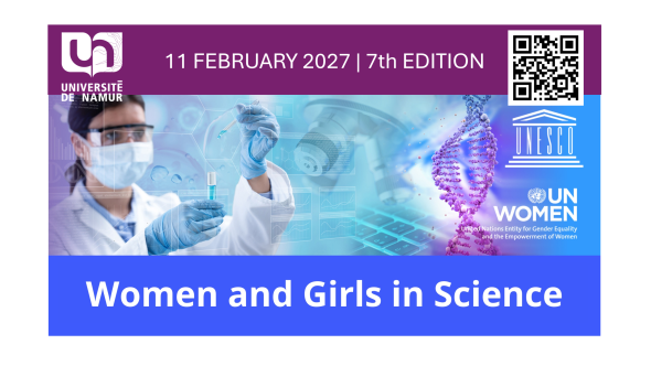
Women in Science - 7th Edition

Event

Women in Science - 7th Edition

Gender and diversity
Science
SDG 5 - Gender equality
11
2027 09:00 - 20:00
Université de Namur, Faculté des sciences, auditoire S01 - rue Grafé, 2 - 5000 Namur
Contact person :  Comité Women in science

This annual event aims at promoting the access of women and girls to science and technology as well as their full and fair participation. It reminds the important role of women in the scientific community and constitutes a great opportunity to encourage girls and young women to participate in the scientific developments.
Women in science - 7th edition

Save the date!

Show your support for equal opportunities for ALL genders in science and technology.

More information to come. 

All events

Management

Frédéric SILVESTRE

Director, Biology Department