Image

Plateforme technologique
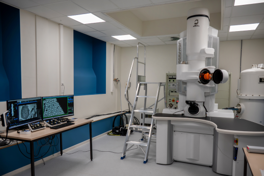
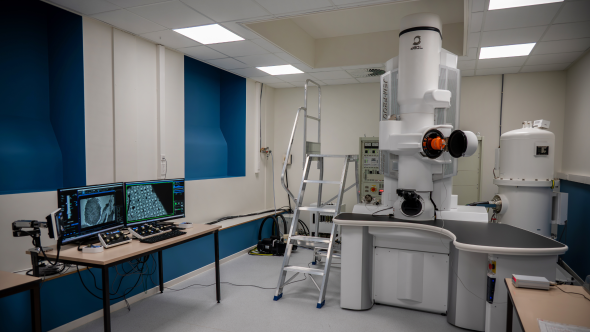
The electron microscopy platform at the University of Namur is open to all researchers wishing to use these techniques to observe or analyze samples, for research or teaching purposes. It also makes its equipment and expertise available to other academic institutions, companies and industries for the analysis of their samples.
Depuis plus de 50 ans, la microscopie électronique est au cœur de la recherche à l’UNamur. Cette technologie utilise un faisceau d’électrons au lieu de la lumière pour illuminer l’échantillon. Les électrons ont une longueur d’onde beaucoup plus courte que celle de la lumière visible, ce qui permet d’atteindre des résolutions nanométriques, voire atomiques.
Au-delà de l’imagerie, la microscopie électronique s’est enrichie de techniques analytiques complémentaires qui permettent d’obtenir des informations chimiques, structurales et spectroscopiques sur les échantillons étudiés. Parmi celles-ci, on retrouve la spectroscopie dispersive en énergie des rayons X (EDS) fournissant des informations complémentaires sur la composition élémentaire de l’échantillon, ou la diffraction d’électrons rétrodiffusés (EBSD), méthode cristallographique utilisée pour déterminer l’orientation des grains, la structure cristalline, et les phases présentes dans un matériau.
Aujourd’hui, la plateforme est équipée de trois microscopes électroniques à transmission et de deux microscopes électroniques à balayage possédant différentes sondes analytiques, ainsi que du matériel nécessaire à la préparation des échantillons. Ces outils font de la microscopie électronique un instrument polyvalent, indispensable dans des domaines aussi variés que la science des matériaux, la biologie cellulaire, la nanotechnologie, ou encore la géologie. Ils permettent non seulement de visualiser les structures mais aussi de comprendre leur composition, leur organisation et leurs propriétés fonctionnelles.
L'équipe de la plateforme de microscopie électronique est constituée d'experts dans les différentes technologies utilisées. Elle comprend un ingénieur, encadrant les analyses réalisées sur les microscopes, et une technicienne chargée de la préparation des échantillons.
Les conditions d'accès et d'utilisation des microscopes et autres appareillages du service par les membres de l'Université sont régis par un règlement. Toute personne souhaitant faire des analyses dans le service est invitée à prendre connaissance de ce règlement.
Pour les membres internes de l'Université de Namur, les procédures, informations et guides d'utilisation sont disponibles sur l'intranet (accès réservé).
Pour les utilisateurs externes (demandes d'analyses), merci de contacter le responsable de service :
Jean-François Colomer - jean-francois.colomer@unamur.be - Tel: +32 (0)81 72 47 08

In the hall of the Faculty of Medicine still sits the first transmission electron microscope, a Philips EM300 used at the Facultés Notre-Dame de la Paix in the 70s. The history of electron microscopy at UNamur began, but the real start was made in 1975 with the acquisition of three more microscopes: two transmission and one scanning. In February 2018, when the technology platforms were created, this department was attached to the MORPH-IM platform, and in April 2024 became the independent "Electron Microscopy" platform, UNamur's 11th technology platform
.
Electron microscopy has become an indispensable technique in many fields of research as varied as materials science (metallurgy, crystallography, etc.) or life sciences (cell biology, medicine, etc.). The principle is to use accelerated electrons, instead of a beam of light as in a conventional photonic microscope, to make much smaller structures observable, right up to atomic resolution.
This technique therefore makes it possible to obtain structural information through imaging, but not only. Thanks to the interaction of electrons with the atoms of the material, other emitted signals can also be analyzed to obtain additional information, for example, on the elemental composition (X-ray analyzer) or crystallography (detector for backscattered electron diffraction) of the sample.
Since 1975 and the initial three electron microscopes, two transmission - Philips EM301 and EM201- and one scanning - JEOL JSM-35 - equipped in 1980 with an X-ray analyzer, electron microscopy at UNamur has evolved in step with the microscopes acquired. A new scanning microscope - a Philips XL20 equipped with an X-ray analyzer - replaced the old one in 1991. Then, in 1999, a new transmission microscope was acquired - a Tecnai10 (FEI)- which was the subject of an article in the newspaper "Le Soir".
The article "Images of the infinitely small shown live" states: "It's not every day that the institution equips itself with a new transmission electron microscope, what's more, the first of its generation in Belgium. (...) The big step of this microscope of a new kind? Its image acquisition process. Whereas previously, images observed on film were fixed using a photographic process, it's now a digital camera coupled to a powerful computer that operates".
Professor Yves Poumay, interviewed at the time, explains, "Some researchers from other universities come to us, not because they don't have equivalent equipment at home, but because it's less accessible or less good"". At UNamur, they "not only provide researchers with a microscope, but also a team of laboratory technicians and an accompanying engineer, which constitutes a rather unusual human framework that is as valuable as the new microscope itself."
The platform's philosophy has not changed, with researchers from all walks of life, but also companies, still and always calling on its expertise.
The modernization of equipment continued in the following years with the acquisition of several scanning microscopes a JEOL JSM 7500F equipped with an X-ray analyzer in 2007 and a JEOL JSM 6010LV in 2012. The latter was very recently modified in 2023 with the acquisition of an X-ray analyzer (SDD QUANTAX, Bruker) and a detector for backscattered electron diffraction (eFlash QUANTAX, Bruker) as part of the inter-university research platform for the energy transition (RRF).
In 2016, a Tecnai20 (FEI), equipped with an X-ray analyzer (SDD QUANTAX, Bruker) mounted in 2021, complements the Tecnai10 for transmission microscopy analyses.
As part of the BIOGREEN technology platform of excellence in 2024, the acquisition of a JEOL JEM F200 cryogenic transmission microscope will add value to the existing fleet of instruments. This new microscope enables the analysis of sensitive materials while minimizing electron beam damage. Thanks to specific preparation methods such as sample vitrification (Leica EM GP2 automatic immersion freezer) coupled with cryo-microscopy, it is thus possible to obtain information on the structure of biological objects (proteins, ribosomes, etc.).
For completeness, the development of electron microscopy has been accompanied by the acquisition of all the equipment required for sample preparation. Some equipment, such as the microtomization equipment, is original (1975), but is in the process of being replaced, while others are more recent, such as the sputtering device (QUORUM QT 150T/ES in 2015).
In 1975, Professor Robert Leloup created the Unité Interfacultaire de Microscopie Électronique, thanks to a substantial budget allocated by the institution. As today, it is located at the corner of rue Grafé and Place du Palais de Justice in the Faculty of Medicine. In 1998, Prof. Yves Poumay took over from Prof. Poumay until 2010, when Dr. Jean-François Colomer became head of the Microscopy Department, and with him a new team was put in place. In 2011, platform engineer Corry Charlier was hired. In 2017, Caroline De Bona joins as a technician. In 2025, the facility becomes part of the FWB's BIOGREEN technology platform of excellence, and a research logistician, Emir Topagolu, joins the team.
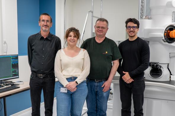
The team: Jean-François Colomer, Caroline De Bona, Corry Charlier and Emir Topaloglu
The "electron microscopy" platform boasts state-of-the-art equipment and remarkable expertise, which it has been sharing for 50 years with all UNamur members who wish to use these microscopic techniques for observation or analysis purposes, from both a research and teaching perspective, whether in materials science or life science. It also makes its expertise available to research organizations from other academic establishments and to companies or industries for the analysis of their samples.
There are two main classes of electron microscope:
This article is taken from the "The Day When" section of Omalius magazine #35 (July 2025).
